Pedantically not phoresy
The life cycle of the ectoparasitic mite Varroa destructor essentially consists of two stages. The first is within the capped cell, where reproduction takes place. The second occurs outside the capped cell when the recently-mated female progeny mites matures while riding around the colony attached to a nurse bee.
Almost without exception this second stage is termed the phoretic phase.
It isn’t.
Phoresy
Phoretic is an adjective of the word phoresy. Phoresy is derived from the French phorésie which, in turn, has its etymological origins in the Ancient Greek word φορησις.
And φορησις means being carried.
Which partly explains why the correct definition of the word phoresy is:
An association between two organisms in which one is carried on the body of the other, without being a parasite [OED]
Phoresy has been in use for about a century, with the word phoretic first being recorded in the Annals of the Entomological Society of America (25:79) in 1932:
It is possible, as suggested by Banks (1915), that such young mites are phoretic, being carried about from place to place on the host’s surfaces.
And, no, they weren’t discussing Varroa.
“Without being a parasite”
These are the critical words in the dictionary definition of phoresy which makes the use of the word phoretic incorrect when referring to mites on nurse bees.
Because mites on nurse bees are feeding – or at least a significant proportion {{1}} of them are.
They are therefore being parasitic and so shouldn’t be described as phoretic.
Om, nom, nom {{2}}
Last week I discussed the recent Samual Ramsey paper presenting studies supporting the feasting of Varroa on the fat body of bees.
In the study they harvested bees from a heavily mite-infested hive and recorded the location on the bee to which the mite was attached.
The majority were attached to the left underside of the abdomen. More specifically, the mite was wedged underneath the third abdominal tergite {{3}}.
What were they doing there? Hiding?
Yes … but let’s have a closer look.
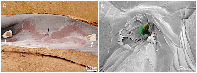
Ramsey and colleagues removed some of the mites and used a scanning electron microscope to examine the attachment point on the bee. Underneath the tergite there is a soft membrane. The imprint of the body of the mite was clearly visible on the membrane.
The footpads of the mite were left attached to the membrane (left image, white arrows), straddling an obvious wound where the mouthparts had pierced the membrane (black arrow). Between them, the inverted W shape is presumably the imprint of the lower carapace of the mite.
The close-up image on the right even shows grooves at the wound site consistent with the mouthparts of the mite.
These mites were feeding.
Extraoral digestion
Varroa belongs to the order (a level of classification) Mesostigmata. Most mesostigmatids feed using a process termed extraoral digestion.
Extraoral digestion has also been termed ‘solid-to-liquid’ feeding. It involves the injection of potent hydrolytic enzymes which digest solid tissue, converting it to a semi-solid that can be easily ingested. It can reduce the time needed to feed and it increases the nutrient density of the consumed food.
If Varroa fed on haemolymph it wouldn’t need to use extraoral digestion. Instead it would need all sorts of adaptations to a high volume, low nutrient diet. Varroa doesn’t have these. It has a simple tube-like gut parts of which lack enzymatic activity … implying that digestion occurs elsewhere.
A picture is worth a thousand words
Do the images of feeding mites support the use of extraoral digestion?
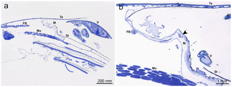
The image above {{4}} shows the cross-section of a Varroa (V), wedged under the tergite (Te), feeding through a hole (arrow in the enlargement on the right) in the membrane (M). The fat body (FB) is immediately underneath the membrane. The scale bar is incorrectly labelled {{5}}.
A close-up of the wound site shows further evidence for extraoral digestion.
Beneath the wound site (C, arrow) are remnants of fat body cells (white arrow) and bacteria (black arrow; of two types, shown in D). A closer look still at the remnants of the fat body (E and F) shows cell nuclear debris (blue arrows) and lipid droplets (red arrows).
These images are entirely consistent with extraoral digestion of fat body tissue by feeding Varroa. The presence of bacteria near the wound suggests that bacterial infection may result from Varroa feeding, possibly further contributing to disease in bees.
So, pedantically it’s not phoresy
So-called phoretic mites, unless they’re on the thorax or head of the bee, are not really phoretic. They are being carried about, but they are also likely feeding. By definition that excludes them from being phoretic.
Instead they are ectoparasites of adult bees.
What are the chances that beekeepers will stop using the term phoretic?
Slim to none I’d predict {{6}}.
And, of course, it doesn’t really matter what the correct term for them is.
What’s more important is that beekeepers remember that it’s at this stage that mites are susceptible to all miticides.
The June gap
But it’s also worth thinking about the potential impact of brood breaks.
During brood breaks all the mites in the colony must be ‘phoretic’.
Generally, the majority of the mites in a hive are in capped cells. Depending upon the stage of the season, the egg-laying rate of the queen and other factors, up to 90% of the mites are associated with developing pupae.
But as the laying rate dwindles more and more mites are released from cells and become ‘phoretic’, unable to find a suitable late-stage larva to infest.
And which bees do the mites associate with?
Nurse bees primarily, for reasons I’ll discuss in the future. But – spoiler alert – one of the reasons is likely to be that they have a larger fat body.
So, a mid-season brood break (e.g. the ‘June gap’) is likely to result in lots more nurse bees becoming both the carriers and the dinner of the mite population.
Some or many of the nurse bee cohort may perish, perhaps from damage to the fat body or from the viruses acquired from the mite. However, bees exhibit phenotypic plasticity, meaning that older bees can revert to being nurse bees when the queen starts laying again.
Late season brood breaks
In late summer mite levels are usually at their highest in the hive. A brood break occurring now will release a very large number of mites to parasitise the adult bee population.
Presumably these mites select the bees best able to support them {{7}}.
And which bees are these? The nurse bees of course. But it’s also worth remembering that there are key physiological similarities between nurse bees and winter bees. Both have low levels of juvenile hormone and high levels of vitellogenin (stored in the fat body).
So I’d bet that the ‘phoretic’ mites during a late season brood break would also preferentially associate with any early-produced winter bees.
Furthermore, once the queen starts laying again – perhaps in early/mid-autumn – the winter bees being produced would be subjected to the double-whammy of high levels of mite infestation and potential damage from ‘phoretic’ mites.
Practical considerations
More work is required to model or actually measure the impact of late season brood breaks, high levels of ‘phoretic’ mites, nurse bee numbers and winter bee development.
Compare two colonies of a similar size with a similar mite load, treated at the same time in early autumn with an appropriate miticide. If one of them experienced a late summer brood break (pre-treatment) and consequent high levels of ‘phoretic’ mites, does this reduce the chances of the colony surviving overwinter?
Who knows? Lots and lots of variables …
Fundamentally, it remains important to treat colonies early enough to protect the winter bee population. You’ve heard this from me before and you’ll hear it again.
However, it’s something to think about and I can see ways in which it might influence the strategy and timing of mite control used. I’ll return to this sometime in the future.
{{1}}: >95% of the mites on adult bees in the recent study by Ramsey et al., 2019.
{{2}}: Where nom is an onomatopoeic adjective, first used by the Cookie Monster on Sesame Street, to indicate the sound of ravenous eating. Nom was defined in 2004, though the Cookie Monster is a whole lot older, and is also precisely the noise Varroa makes … if you listen very, very carefully.
{{3}}: The sclerotized plate over the abdominal segment.
{{4}}: It’s a longitudinal section through the bee. The head end is to the left side of the image, but it’s only showing a narrow slice of the abdomen.
{{5}}: If it isn’t the Varroa is about 2 feet long!
{{6}}: And I’ll struggle as well … I’m going to try and use quotes to enclose the word ‘phoretic’ in the remainder of this and future posts.
{{7}}: Evolution tends to arrange these sort of things rather neatly.


Join the discussion ...